Hair Mineral Test - FAQ, Facts and Information
Hair mineral analysis identifies chronic toxic metal exposure and mineral imbalance, factors that can lead to a suppressed immune system and a degeneration of the ability to self-heal. Hair mineral analysis shows you any mineral deficiency and toxic metal you have, giving you the opportunity to rebalance your body holistically. Prevention is always better than cure.
The Joint United Nation Food and Agriculture Organization and the World Health Organization Expert Committee for Food Additives and Contaminants 61st meeting recognised that hair test is the preferred method of identifying chronic mercury exposure, whilst a blood test is appropriate for acute exposure. Exposure to mercury is low dose build up over the long-term. Therefore, hair is a more appropriate specimen sample.
The mercury level in hair is 250 –300 times higher than that of blood. Hair can reflect even low-levels of mercury poisoning more accurately than blood.
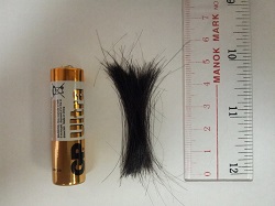
The amount of hair needed is 0.25g (about the weight of 8 staples). We will collect a small
bundle of hair from6-8 different areas from the back of the head.


We do not recommend clients who have dyed their hair within the past 30 days to do this test. Most shampoo will not affect the result, except for anti-dandruff formulas containing selenium which will affect the reading of selenium only.
You can still do this test because armpit hair, pubic hair or nails can be used as substitutes.
The turnaround time is about 10-14 days.
Whether a re-test is needed or not depends on the results of the first test. A re-test is usually performed 9 months after the first test.
A unique window on metabolic activity HTMA is a precise analytical test that assesses the mineral composition of the hair, providing a sensitive indicator of the long-term effects of diet, environment, stress and toxic metal exposure.
As one of the body's most metabolically active tissues, hair is exposed to blood, lymph and intracellular fluids. As the cluster of matrix cells, which form a hair follicle, reach the surface of the skin the outer layers harden locking in the trace elements that accumulated during growth and providing, in effect, a biochemical blueprint. Clinical studies and a wealth of scientific literature have shown that when sampled and analysed correctly, hair analysis can give an accurate indication of nutrient mineral excesses, deficiencies, biochemical imbalances and toxic metal accumulation.
Since hormonal activity is well known to affect the metabolic utilization of certain trace nutrients, HTMA is now used widely by medical practitioners to aid in specifying a patient's metabolic type. The correlation of over 300,000 tissue mineral patterns with specific physical and biochemical characteristics has enabled Dr Watts and his team at Trace Elements to identify eight distinct metabolic categories. These are fast and slow metabolic types each with their four sub-types, which are associated with the various stages of stress, whether acute or chronic in nature.
HTMA reveals cellular metabolic activity unattainable through most other tests. It is a screening tool applicable to both assessments of health status and toxic metal accumulation, while also serving as an invaluable diagnostic aid. If a patient is suffering from an illness or syndrome which cannot be identified through routine clinical tests, hair analysis can help to pin point a possible metabolic disturbance and help the clinician choose the appropriate course of treatment. At the very least, hair analysis should be used once a year to evaluate toxic metal exposure and accumulation.
Although blood and serum give an insight into the transport of certain elements around the body, they are not accurate indicators of the actual storage of elements within the cells. Monitoring trace elements in urine can only measure components which are being excreted, not absorbed. Serum concentrations may fluctuate with the manner in which the sample is taken, emotional changes, the time of day the blood is drawn, or foods eaten prior to taking the sample.
For example: Eating bananas before a blood test will cause a rise in blood potassium, but blood re-tested the following day may show blood potassium to be normal or even low.
Similarly, calcium levels assessed by a blood test do not truly reflect mineral status in the body and calcium loss from the bones can be quite advanced before significant changes in blood calcium levels are detected.
Iron deficiency symptoms can be present in hair long before low levels are seen in serum.
Thirty to forty days following an acute exposure, elevated serum levels of lead may be undetectable. This is due to the body's removing the lead from serum as a protective measure and depositing them into tissues such as the liver, bones, teeth and hair.
- Hair samples can be collected easily and painlessly using non-invasive techniques.
- Hair is less susceptible to the homeostatic mechanisms that quickly affect trace element levels in the blood. It is therefore a more reliable indicator of nutritional status or imbalance.
- Concentrations of most elements in the hair are up to ten times higher than levels found in blood and other tissues.
- Trace element levels found in hair tissue represent time-weighted exposure (providing a record of past and present nutritional status) making it more useful for epidemiological and nutritional studies.
Hair is the tissue of choice for determining toxic metal exposure by the US Environmental Protection and it is used for this purpose worldwide.
Minerals are the spark plugs in the chemistry of life. Their functions range from providing structural support in the formation of bones and teeth, to nerve conduction, muscle contraction, the production of enzyme and hormones such as insulin and the maintenance of the acid-base balance in the body. Research is now recognising their importance in the healthy functioning of the immune system, the fight against ageing and the prevention of chronic degenerative diseases.
Minerals and vitamins co-exist in a delicate balance, which is influenced by the activity of the endocrinal glands. The key to understanding the effect of nutrients is to understand these interrelationships. For example, a deficiency of vitamin C leads to a build up of copper to often toxic levels. Too much copper can cause a deficiency of iron, selenium and potassium. Conversely, excess vitamin C can result in copper deficiency leading to iron retention in the body.
Anemia:
Iron deficiency is suprisingly common. It frequently occurs without symptoms of anemia and often when blood serum iron falls in the "normal" range. This is because blood maintains serum iron at the expense of tissue. Symptoms include fatigue, abnormalities of the gastro-intestinal tract and learning difficulties in children.
Pregnancy:
Iron supplements are routinely prescribed to pregnant women with little regard to their depleting effect on a woman's zinc levels. Zinc deficiency has been linked closely to poor pregnancy outcome and congenital malformations.
Osteoporosis:
Over 30 mechanisms have been linked to porous and brittle bones, for example too much calcium can actually cause osteoporosis in bone tissue lacking magnesium, phosphorous, zinc, and copper. Typically, the body deposits magnesium on the surface of the bones, thus a relative deficiency will produce a thinning of the bone cortex making them more susceptible to breaks.
Diabetes:
Calcium is a factor in the release of insulin. Chromium is a well-known component of glucose tolerance factor (GTF) and acts by increasing insulin efficiency. Excessive quantities of glucose and insulin however are known to increase chromium excretion. Multiple Sclerosis: The symptoms of mercury poisoning can mimic those of multiple sclerosis and amyotrophic lateral sclerosis (Lou Gehrig's disease). Furthermore copper, which is required for the mylelination of nerves is often severly lacking in patients suffering from MS or Parkinson's disease.
Impaired Mental Function:
The brain stores trace elements in various sectors. An abnormal concentration or imbalance among these minerals can affect psychological functions, including emotions, memory, perception, learning and behavior. Excessive levels of calcium are a documented cause of depression and elevated copper levels are closely associated with post partum depression, PMS and dyslexia.
Diet:
 A major factor in contributing to a mineral imbalance is improper eating habits. Excessive intake of refined and processed foods, alcohol and fad diets can all lead to poor mineral nutrition. Even the nutrient content of a "healthy" diet can be inadequate, depending on the soil in which the food was grown or the method in which it was prepared. Example: Inadequate protein intake, excess sugar and high vitamin D intake can result in decreased calcium utilization by the body leading to reduced energy levels, joint stiffness and changes in skin and hair texture.
Stress:
Stress either physical or emotional, can lead to mineral imbalances. Certain nutrients such as the mineral zinc and the B-complex vitamins are lost in greater quantities due to increased stress. Nutrient absorption can decrease quite significantly when the body is under stress.
Medications:
Medications can deplete the body store of nutrient minerals or increase the levels of toxic metals. The well-known effect of diuretics include not only sodium loss but, in many cases, a potassium and magnesium loss. Antacids, aspirin, and oral contraceptive agents can lead to vitamin and mineral deficiencies as well as toxic metal excesses.
Pollution:
 Toxic metals such as lead, mercury, and cadmium can interfere with mineral absorption and increase mineral excretion. From adolescence to adulthood the average person is continually exposed to a variety of toxic metal sources such as cigarette smoke (cadmium), cookware (copper and aluminum), hair dyes and cosmetics (lead), hydrogenated oils (nickel), antiperspirants (aluminum), and contaminated seafood (mercury).
Nutritional Supplements:
Vitamin and mineral supplements can also lead to mineral imbalances. Calcium absorption is decreased in the presence of phosphorous. Vitamin D enhances calcium absorption, but in excess amounts, can produce a magnesium deficiency. Inherited Patterns: A predisposition towards mineral deficiencies, excesses and imbalances can be inherited from the parents.

